
Highlight
In Situ Synthesis of Calcium Phosphates in Block Copolymer Hydrogels
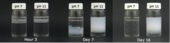
Images of Ca2+ diffusion in to phosphate containing hydrogel at 3 hours, 7 days and 16 days after calcium addition.
Achievement/Results
Polymeric composites have recently gained a great deal of attention for their use in biomaterial and tissue engineering applications. Combining two or more, sometimes very dissimilar, components gives composites properties unattainable using only a single constituent part. Polymer composites containing calcium phosphate minerals have been especially popular due to their biocompatibility and in vivo resorption capacity. Physical properties of calcium phosphate composites can additionally be used as a predictor for their efficacy in tissue engineering applications. Mineral phase, size and morphology can have a great effect on in vivo dissolution rate and the overall composite’s mechanical response, which in turn should be similar to that of native tissue to prevent an adverse reaction. An IGERT student in Prof. Surita Bhatia’s group in Chemical Engineering, Mr. David Griffin, focuses on the in situ formation and characterization of calcium phosphate composites in self-assembling block copolymer hydrogels. The approach is highly interdisciplinary and combines synthetic techniques from inorganic chemistry, structural and rheological characterization techniques from polymeric materials, and knowledge of biological structure and function. Initial work has concentrated on gels of poly(ethylene oxide)-poly(propylene oxide)-poly(ethylene oxide) (PEO-PPO-PEO), specifically Pluronic® F127. Not only has this polymer been shown to contain biocompatible segments, but it is also readily available in large quantities and has been thoroughly characterized.
Hydrogel mineralization is carried out by first dissolving a phosphate precursor (phosphoric acid) and polymer in water at low temperature (4 °C). The addition of sodium hydroxide to this solution allows us to control the initial pH of our samples as well. By removing solutions to room temperature the polymer undergoes a spontaneous thermodynamic transition to a micellular structure and forms an interconnected, physically-linked hydrogel. A small volume of calcium chloride solution is then pipetted on top of the gel and allowed to diffuse and react with the phosphate precursor. By utilizing this method we are able to induce in situ nucleation and growth of calcium phosphate minerals in the gel phase.
Initial examination of our samples showed that pH plays an important role in the calcium phosphate formation. Various calcium phosphate phases exhibit substantially different solubilities dependent on pH which is evident in the appearance of the composite hydrogels. Composites formed in hydrogels with an initial pH 7 are transparent, like the neat gel, with dispersed large crystallites on the size scale of millimeters. Composites formed at pH 11, however, are opaque and milky white in appearance indicative of a homogeneously distributed, smaller mineral particle.
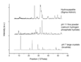
X-ray diffraction (XRD) analysis on the composite minerals was able to identify the calcium phosphate phases present in our samples. At near-physiological pH the mineral is a highly crystalline brushite phase, whereas at basic conditions we obtained calcium hydrogen phosphate hydrate. Furthermore, we desired to compare our composite minerals to that of commercially available synthetic hydroxyapatite (HAp). Hydroxyapatite was chosen due to its chemical and structural similarity to the natural inorganic component of bone tissue. Of particular note was that the crystallographic peaks in the diffraction pattern of the pH 11 mineral appeared in d-space ranges similar to that of hydroxyapatite. Peak broadening of the calcium hydrogen phosphate hydrate also suggested a nanocrystalline mineral component.
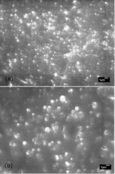
Scanning electron microscope (SEM) images were taken of the pH 11 mineral to determine size and morphology. These confirmed previous results from light scattering that the composite was indeed composed of nanoscopic mineral particles. Additionally, the calcium phosphate particles appeared disk-like in shape, similar to apatite particles in the mineralized collagen of bone. When compared with commercial HAp, the pH 11 mineral was strikingly similar in appearance.
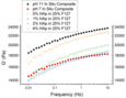
Yet another important property of our composite hydrogels was discovered after examining their mechanical response to stress. Rheological measurements showed that the in situ composites exhibited elastic moduli in the 15-20 kPa range. What was outstanding, however, was how these samples compared to F127/hydroxyapatite composites produced by conventional mechanical mixing. While the in situ composite manufactured at basic conditions had a solids content of approximately 1%, its elastic moduli was greater than conventional composites with a mineral load four times higher.
Further progress in this investigation is likely to lead in many new and exciting directions. Techniques in synthetic polymer chemistry make it possible to alter the copolymer functionality and examine polymer/mineral interactions in the composite, and collaborations along these lines are being discussed with the group of Prof. Gregory Tew in Polymer Science and Engineering. In addition, cellular biology approaches can be applied to inspect mammalian cell response to composites in vitro, and discussions are underway with the groups of Prof. Janice Telfer (Veterinary and Animal Science) and Prof. Susan Roberts (Chemical Engineering) to initiate this work. Our novel approaches to biomaterials design have the potential to impact repair of diseased and damaged bone and cartilage and thus has important societal benefits.
Address Goals
The primary goal is addressed in that the research addresses an area of great opportunity and potential benefit; that is, the design of improved biomimetic materials that may have applications in treatment of bone and cartilage disorders. The work being performed is at the frontier of materials research and combines knowledge from materials science, synthetic chemistry, and bioengineering.
The secondary goal is addressed in that the graduate student involved in the work is gaining an appreciation of how interdisciplinary research is performed, contributing to a well-trained workforce. The particular student involved in the work, David Griffin, has also been active in mentoring undergraduate researchers and giving laboratory tours and demonstrations to visiting high school students, expanding the scientific literacy of undregraduates and the public.