

Highlight
Microbead Array Platforms for High Throughput Screening
Arraying of Microbeads Using Microfluidic Entrapment
Achievement/Results
Background The proteins of a cell, a tissue, or a complex organism orchestrate biological activity by binding. Proteins bind to other proteins and as well to small molecules like peptides, carbohydrates, or nucleic acids. The binding interactions of proteins which direct activity are interconnected in a complex network, and mapping the network leads to a scientific understanding of the contributing roles of each protein. Platforms which allow large numbers of proteins and protein-binding biomolecules to be rapidly screened against one another in order to identify binding partners not only allows this complex web to be deciphered, but they can also be used as technological tools for the advancement of human health.
The standard platform for assaying the interactions of large numbers of proteins or protein-binding biomolecules are protein microarrays, which are surfaces consisting of robotically-dispensed individual spots of capture or probe biomolecules (adsorbed or covalently linked to the surface), hundreds of microns in diameter, which are arranged in a spatially indexed grid pattern on a surface. An analyte sample containing potential binding targets to the array of displayed capture molecules is incubated over the surface, and binding events are identified, usually by fluorescently labeling the targets (or the products of an enzyme linked assay), spatially scanning the array for the luminescence and finally matching the probe to the target.
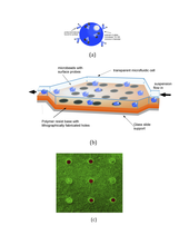
Achievement A new particle-based microarray platform has been developed to enable high througput screening of the binding interactions of biomolecules. In this platform, microbeads (glass or polymer particles tens of microns in diameter) are used as hosts for the biomolecules (the probes) whose binding interactions are to be screened against a particular biomolecule of interest (the target). The probe molecules are chemically linked or adsorbed to the surface of the bead, with each bead having only one kind of probe molecule linked to its surface. (see image) In order to implement a screening platform, microbeads, each displaying a different probe, are arrayed on a surface in fixed locations which are indexed so that the probe at the location is known. A microfluidics cell is prepared which consists of a transparent channel which is rectangular in cross section, with a height approximately equal to that of the beads and a width and length of a few millimeters. At the bottom of the channel are fabricated an array of traps designed to capture – as an obstacle course – the microbeads as they flow in an aqueous suspension through the cell. One type of entrapment course which is being studied consists of wells patterned at the bottom of the channel to capture the microbeads as they drop by gravity into the wells.
The microfluidic obstacle courses are filled with microbeads containing different probes sequentially: Beads with one particular probe are flowed, trapped and indexed (by inspecting the array with an optical microscope), and the process is then repeated to assemble a multiplexed platform displaying a number of probes. After the trapping array is filled, the target molecule can be flowed through the array, and binding events identified by fluorescently labeling the target, and examining the array for fluorescing microbead locations
Using this microbead design for a high throughput screening platform, the microfluidic capture of the particles in wells has been demonstrated. The microfluidic cells are constructed using soft lithography techniques in which the well surface (the bottom of the microfuidic cell) is first fabricated out of polydimethylsiloxane (PDMS) by pouring the liquid pre-polymer over a mold of pillars, curing the polymer and peeling the polymer from the mold. The top of the cell, which consists of a rectangular cavity (50 microns in depth, millimeters in width and length) inscribed on one face of a PDMS slab, is formed next by casting off of a second mold. Inlet and exit ports at opposite sides of the top of the cell are punched in the PDMS into the cavity to allow for the deposition of the microbeads into the cell. The top and bottom of the cell are adhered, and the ports are connected to tubing with the tubing for the inlet connected to a syringe pump. The microbeads, suspended in water, are pumped through the inlet port, flow across the surface and exit through the outlet port. As they flow across the well surface they drop into the wells due to gravity (the cell is orientated horizontally).
The microbeads are monodisperse glass particles 40 microns in diameter which have been functionalized with a fluorescein dye on their surfaces in order to view the beads by their surface fluorescence once the dye is excited by illumination at its excitation wavelength. The depth and diameter of the wells are slightly larger (50 microns) than the diameter of the particles. The glass beads are heavy enough to be readily deposited in the wells at relatively low flow rates, and beads remaining on the surface but not in wells are easily cleared without the entrapped beads being driven out of the wells. The surface of the microbeads and the inside surfaces of the microchannel have been functionalized with polyethylene glycol (PEG) oligomers in order to reduce nonspecific adsorption of biomolecules and allow the microbeads to roll easily over the bottom surface. The image shows fluorescent and phase contrast images of labeled microbeads partially filling an array obtained using a laser scanning confocal microscope. The images demonstrate that microfluidic assembly through an obstacle course can be used to array microbeads.
Further results have been obtained on arraying two kinds of microbeads, distinguished by using different fluorescent labels on the bead surfaces. The viability of the platform as a diagnostic device for undertaking the screening of biological interactions has also been demonstrated using the selective binding of neutravidan to biotin.
Address Goals
Transformative Nature of the Achievement This bead based platform for screening the interactions of biomolecules can represent a major advance in high throughput screening, particularly because of its miniaturized scale. The current microarray technologies use robotic dispensing to form arrays of spots of probe molecules are tupcally several hundred microns in diameter. The microbead platform, which uses particles as hosts for the probes, and a simple fluidic cell and hydrodynamic capture to form the array, implement a much easier method for arraying and a size for the probe location which is at least an order of magnitude smaller. The miniaturization allows for a greater throughput and a reduced reagent consumption during the assaying. In addition, the microbead platform enables the display of membrane protein receptors. These class of receptors are the most important for regulating activity on the cellular scale, and are the primary focus in drug discovery efforts as developing drugs to bind to membrane protein receptors can lead to strong therapeutic effects. To retain their binding ability, membrane proteins need to be sequestered in their native lipid bilayer environment of a cell membrane. As such they cannot be directly spotted onto a surface. However, microbeads can be coated with lipid biilayers, and the bilayers then sequestered with the membrane proteins. In this way arrays of membrane proteins can be constructed, forming platforms for screens of these important receptors which were not possible with the current technology.
Societal Benefits The societal benefits revolve around the applications of the microbead biomolecular screening platform. The platform arrays many different probe biomolecules on microbeads to be screened against other target biomolecules to identify binding partners. Screening arrays can be used to identify drug molecules which conjugate to a particular, intended receptor and as a point-of-care diagnostic device to screen for disease markers. The ease with which the microbead array can be assembled will enable a wider accessibility compared to other microarray screening technologies which involve more complicated fabrications. And the fact that the microbead platform can easily be adopted to display membrane receptors by using lipobeads should provide use in important areas such as drug discovery where drug candidates target and need to be screened against a wide palette of membrane receptors.
