
Highlight
Computational Analysis Reveals Increased Blood Deposition Following Repeated Mild Traumatic Brain Injury
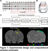
Figure 1: Experimental design and computational analysis schematic.
Achievement/Results
NSF-funded researchers at the University of California, Riverside have demonstrated for the first time that the evolution of tissue damage caused by repeated mild traumatic brain injury (mTBI; or concussion) is dependent on the time between injuries. The NSF IGERT in Video Bioinformatics at the University of California, Riverside (UCR) brought together an interdisciplinary team of engineers and biologists that developed a novel computational method for magnetic resonance imaging (MRI) analysis. These findings provide additional insight into the ongoing debate of “return to play” definitions in athletes and resuming military duties after sustaining a mild injury. Previously, this study was not possible by processing the magnetic resonance (MR) images by hand. It has been enabled with a new algorithm that efficiently quantifies blood and edema volumes—important for analysis of mTBI—for accurate analysis of neuropathology. With this algorithm, the temporal development of cortical damage following two mTBIs (induced to opposite hemispheres) at 3 (rmTBI 3d) and 7 (rmTBI 7d) days apart was examined (Fig. 1A, B). Voxels containing blood, edema, normal appearing brain and noise are identified from user-defined regions of abnormal cortical tissue (Figure 1C, D).
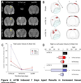
This study has two novel findings: (1) abnormal tissue volumes were significantly increased in animals receiving mTBIs 7d apart, particularly at the site of second injury (left; Figure 2), and 2) computationally identified tissue abnormalities revealed increased blood deposition in animals receiving mTBIs 7d apart, but increased edema in animals receiving mTBIs 3d apart (Figure 3). These results suggest that injuries induced 7 days apart result in worsened outcomes, such as increased blood which is particularly toxic to brain tissue. Experimentally, we have developed a new computational method for quantitative MRI analysis, which has not been done before in models of mild injury
Performers:
Virginia Donovan; Anthony Bianchi; Richard Hartman; Bir Bhanu; Monica J Carson; Andre Obenaus
References:
Virginia Donovan; Anthony Bianchi; Richard Hartman; Bir Bhanu; Monica J Carson; Andre Obenaus, “Computational Analysis Reveals Increased Blood Deposition Following Repeated Mild Traumatic Brain Injury,” Submitted to NeuroImage.>
Anthony Bianchi, Bir Bhanu, Virginia Donovan and Andre Obenaus, “Contextual and Visual Modeling for Detection of Mild Traumatic Brain Injury in MRI,” International Conference on Image Processing, 2012.
Address Goals
The activity contributes to the state of the art. Mild Traumatic Brain Injury is a common topic of discussion in the news.
Virginia Donovan (PhD student in Cell, Molecular and Development Biology) and Anthony Bianchi (Electrical Engineering) are IGERT fellows who were trained and worked on this interdisciplinary project.