

Highlight Photo
IDENTIFYING THE GEOMETRIC TRIGGERS FOR CELL STEMNESS
GEOMETRIC MICROCUES FOR CELL STEMNESS
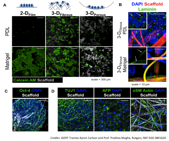
CAPTION: Figure 1 (A) hESCs form viable colonies within PDL-treated fibrous scaffolds only, in contrast to Matrigel-treated substrates, which support hESCs in 2-D and multiple 3-D configurations. (B) hESCs deposit ECM protein laminin in intercellular spaces on PDL-treated scaffolds, in contrast to Matrigel-treated scaffolds, where the only laminin present is that deposited on fibers during pretreatment. © hESCs under maintenance conditions maintain expression of self-renewal marker Oct4 after 14 days of culture. (D) hESCs can be differentiated within PDL-treated fibrous scaffolds to TUJ1+ neuronal cells, AFP+ hepatic cells, and smooth muscle actin+ smooth muscle cells in situ by treatment with soluble factors.
Credits: Aaron Carlson and Prabhas Moghe, Rutgers
