

Highlight Photo
Fluorescent Wide-Field Imaging of Nanoparticles In Vivo
In vivo images of fluorescent nanoparticle spacers
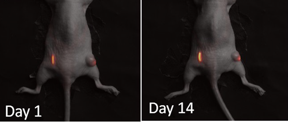
In vivo imaging of spacers inserted into the left flank and a tumor in the right flank. Image from Day 1 after insertion (left) and day 14 (right) show fluorescence in tissue from the NP coated spacer.
Credits: NSF IGERT trainee Stacey Markovic and advisor, Prof. Mark Niedre from IGERT Nanomedicine Science and Technology Program at Northeastern University.
